

Medical imaging
Medical imaging is the group of techniques that produce pictures of the inside of the body, such as X-ray radiography, computed tomography (CT), magnetic resonance imaging (MRI), ultrasound and nuclear medicine scans, so that clinicians can detect, diagnose and monitor disease and guide treatment.[1][2][3][4][5] Radiology accounts for most of the artificial intelligence medical devices authorized in the United States: in the September 2026 version of the U.S. Food and Drug Administration's (FDA) list of AI-enabled medical devices, 1,230 of the 1,614 listed devices (about 76%) had radiology as their lead review panel.[6] Since the mid-2010s, research in the field has relied mainly on deep learning, in particular convolutional neural networks, for image classification, detection, segmentation and registration.[7] Large randomized and real-world screening studies have reported gains in cancer detection, while systematic reviews and validation studies have documented weak study designs, poor generalization to new hospitals, and bias against under-served patients.[8][9][10][11][12]
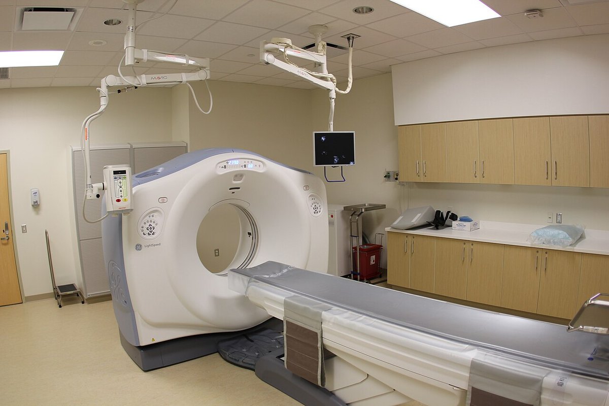
Imaging modalities
Wilhelm Röntgen discovered X-rays in 1895 while studying cathode radiation, and received the 1901 Nobel Prize in Physics for the discovery.[13] The modalities in routine clinical use today differ in the physics they rely on, whether they expose the patient to ionizing radiation, and the tissues they show best.
| Modality | How the image is formed | Ionizing radiation | Typical uses |
|---|---|---|---|
| Radiography and mammography | X-rays pass through the body and strike a detector on the other side, forming a shadow-like image[1] | Yes[1] | Fractures, lung infections, kidney stones, breast cancer screening[1] |
| Computed tomography (CT) | An X-ray source rotates around the patient; a computer builds cross-sectional slices that can be stacked into a 3D volume[2] | Yes[2] | Head injury, stroke and bleeding, lung tumors and clots, abdominal lesions, complex fractures[2] |
| Magnetic resonance imaging (MRI) | A strong magnet aligns protons in the body's water; radiofrequency pulses knock them out of alignment and the scanner detects the energy released as they realign[3] | No[3] | Brain, spinal cord, nerves, muscles, ligaments and tendons; tumors and aneurysms[3] |
| Ultrasound | A transducer emits high-frequency sound and builds images from the returning echoes[4] | No[4] | Pregnancy monitoring, heart, blood vessels, abdominal organs, needle guidance[4] |
| Nuclear medicine (PET, SPECT) | Injected radiotracers emit gamma rays (SPECT) or positrons whose annihilation with electrons produces pairs of gamma rays (PET); detectors record the gamma rays and a computer builds 3D images[5] | Radioactive tracer[5] | Cancer detection and staging, heart and brain imaging, bone disorders[5] |
Nearly all radiology, cardiology and radiotherapy equipment uses DICOM (Digital Imaging and Communications in Medicine), the international standard for medical images and related information. It was first published in 1993, the National Electrical Manufacturers Association (NEMA) serves as its secretariat, and the International Organization for Standardization recognizes it as ISO 12052.[14] Images and their radiology reports accumulate in hospitals' picture archiving and communication systems (PACS); the authors of the ChestX-ray8 dataset presented their work as a way to put such hospital-scale archives to use in deep learning, mining disease labels from the accompanying reports.[15]
Computer analysis before deep learning
Software that reads medical images predates deep learning by decades. The FDA's AI-enabled device list begins in 1995 with two pathology devices, the AutoPap 300 QC automatic Pap screener and the PAPNET Testing System; its first radiology entry is the M1000 ImageChecker, authorized on 26 June 1998 and now listed under Hologic.[6] After the FDA approved computer-aided detection (CAD) for mammography in 1998 and the Centers for Medicare and Medicaid Services raised payment for it in 2002, CAD spread quickly through U.S. breast screening.[16]
Evidence that it improved accuracy was sparse. A 2015 study in JAMA Internal Medicine compared 495,818 digital screening mammograms read with CAD against 129,807 read without it, from 323,973 women, interpreted by 271 radiologists at 66 facilities of the Breast Cancer Surveillance Consortium between 2003 and 2009.[16] Screening performance did not improve on any measure: sensitivity was 85.3% with CAD and 87.3% without, and the cancer detection rate was 4.1 per 1,000 women in both groups. Among radiologists who read both ways, sensitivity was lower with CAD.[16] The authors noted that CAD cost more than $400 million a year and concluded that insurers were paying more for it "with no established benefit to women".[16]
Deep learning in medical imaging
A 2017 survey in Medical Image Analysis by Geert Litjens and colleagues summarized more than 300 contributions, most from the previous year, and described convolutional networks as having "rapidly become a methodology of choice for analyzing medical images".[7] The survey grouped the work into image classification, object detection, segmentation, registration and other tasks, across neuro, retinal, pulmonary, digital pathology, breast, cardiac, abdominal and musculoskeletal imaging.[7]
Landmark diagnostic studies
Several studies published between 2016 and 2020 compared deep learning models directly with medical specialists.
| Year | Study | Task | Data | Reported result |
|---|---|---|---|---|
| 2016 | Gulshan et al., JAMA[17] | Referable diabetic retinopathy from retinal photographs | 128,175 training images graded by 54 ophthalmologists and residents | AUC 0.991 on EyePACS-1 and 0.990 on Messidor-2[17] |
| 2017 | Esteva et al., Nature[18] | Skin cancer from clinical photographs | 129,450 images covering 2,032 diseases | Performance on par with 21 board-certified dermatologists on two biopsy-proven tasks[18] |
| 2017 | Rajpurkar et al., CheXNet[19] | Pneumonia on chest X-rays | ChestX-ray14, over 100,000 frontal images | Exceeded average radiologist performance on the F1 metric, against four radiologists[19] |
| 2020 | McKinney et al., Nature[20] | Breast cancer on screening mammograms | Large U.K. and U.S. datasets | Fewer false positives and false negatives; AUC 11.5 percentage points above the average of six radiologists[20] |
In the Gulshan study, the algorithm was evaluated at two operating points; at the high-specificity point it reached 90.3% sensitivity and 98.1% specificity on EyePACS-1.[17] The McKinney study reported absolute reductions of 5.7% (U.S.) and 1.2% (U.K.) in false positives and 9.4% and 2.7% in false negatives, and estimated in a simulation of U.K. double reading that the system could cut the second reader's workload by 88% while maintaining non-inferior performance; the authors presented the work as groundwork for clinical trials.[20] The AUC figures in these papers are the area under the curve of the receiver operating characteristic.
Segmentation
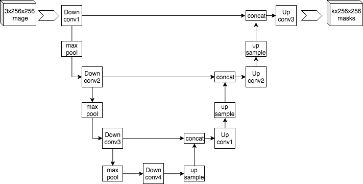
Segmentation delineates organs, tumors and other structures within an image; the developers of MedSAM describe it as a critical part of clinical practice that supports diagnosis, treatment planning and disease monitoring.[21] The U-Net architecture, presented by Olaf Ronneberger, Philipp Fischer and Thomas Brox at the MICCAI 2015 conference, was designed specifically for biomedical image segmentation.[22] It pairs a contracting path that captures context with a symmetric expanding path for precise localization, relies heavily on data augmentation to learn from few annotated images, and won the ISBI 2015 cell tracking challenge in two categories by a large margin.[22]
Because a specialized segmentation pipeline depends heavily on the properties of each dataset and on the available hardware, a group at the German Cancer Research Center built nnU-Net, a method that configures its own preprocessing, network architecture, training and post-processing for any new task.[23] Published in Nature Methods in December 2020, it outperformed most existing approaches, including specialized ones, on 23 public datasets from international segmentation competitions without manual intervention.[23] The Medical Segmentation Decathlon, a challenge that asked algorithms to handle many tasks and modalities at once, reached a related conclusion: state-of-the-art methods generalized well when retrained on unseen tasks, and the organizers wrote that training accurate segmentation models had become "commoditized" for scientists without AI expertise.[24]
Public datasets
The CheXpert authors credit large labeled datasets with driving deep learning to expert-level performance on many medical imaging tasks.[25] In chest radiography, several public datasets were labeled automatically from radiology reports rather than by hand.
| Dataset | Release | Contents | Labels |
|---|---|---|---|
| ChestX-ray8 / ChestX-ray14 (NIH Clinical Center) | 2017 | 108,948 frontal X-rays of 32,717 patients in the first release[15]; 112,120 images from 30,805 patients in the NIH set used by Zech et al.[11] | Disease labels text-mined from reports with natural language processing[15] |
| CheXpert | 2019 | 224,316 chest radiographs of 65,240 patients[25] | 14 observations, with explicit "uncertain" labels[25] |
| MIMIC-CXR (Beth Israel Deaconess Medical Center) | 2019 | 377,110 images from 227,835 studies of 65,379 patients[26] | De-identified free-text radiology reports[26] |
Using the NIH set together with radiographs from two other hospital systems, Zech and colleagues showed in 2018 that a model can learn to recognize the hospital an image came from, not only the disease.[11]
Image reconstruction
Deep learning is also used in image reconstruction, the step in which a scanner's raw measurements are turned into images. The FDA list includes GE's "Deep Learning Image Reconstruction" for CT, first entered in April 2019, Canon's AiCE reconstruction for MRI systems, GE's AIR Recon DL, and Subtle Medical's SubtleMR and SubtlePET software, among other entries.[6]
Foundation models
More recent work applies foundation models, large models meant to be reused across many tasks with little task-specific data, to medical images.[27] The Segment Anything Model (SAM), a promptable segmentation model described in an April 2023 paper, was built with a dataset of more than one billion masks on 11 million images.[28] MedSAM, a foundation model for universal medical image segmentation published in Nature Communications in January 2024, was developed on 1,570,263 image-mask pairs from 10 imaging modalities and more than 30 cancer types, and its authors reported better accuracy and robustness than modality-specific specialist models across 86 internal and 60 external validation tasks.[21] Google's MedGemma, described in a July 2025 technical report, is a family of vision-language models built on Gemma 3 4B and 27B; the report includes chest X-ray finding classification among its evaluations and introduces MedSigLIP, a medically tuned image encoder.[27] A Congressional Research Service report of 10 June 2026 said that, to that date, the FDA did not appear to have authorized any generative AI-enabled device for marketing.[29]
Regulation
United States
In the United States, AI-enabled devices, often software as a medical device, are regulated like other devices under a risk-based scheme of Classes I to III. Most AI-enabled devices have been placed in Class II (moderate risk) and reached the market through 510(k) premarket notification, which requires a determination that the device is "substantially equivalent" to a legally marketed device; novel low- or moderate-risk devices can use the De Novo pathway, and some high-risk Class III devices need premarket approval (PMA) with clinical evidence.[29]
The FDA publishes a list of AI-enabled devices authorized for marketing. The agency describes it as "not a comprehensive resource": devices are included mainly when AI-related terms appear in their authorization summaries or classification, and the FDA has said it will explore ways to tag devices that incorporate foundation models, including large language models.[6] The version current as of 4 September 2026 listed 1,614 devices with decisions through 29 June 2026. Radiology was the lead review panel for 1,230 of them, followed by cardiovascular (154) and neurology (73).[6] Authorizations by year, counted from that version of the list:[6]
| Decision year | Devices on the list | Radiology lead panel |
|---|---|---|
| 2016 | 18 | 11 |
| 2017 | 27 | 16 |
| 2018 | 65 | 40 |
| 2019 | 80 | 51 |
| 2020 | 114 | 99 |
| 2021 | 129 | 110 |
| 2022 | 162 | 138 |
| 2023 | 226 | 178 |
| 2024 | 235 | 183 |
| 2025 | 335 | 254 |
| 2026 (to 29 June) | 181 | 135 |
Two De Novo authorizations in 2018 set regulatory precedents. Viz.ai's ContaCT, authorized on 13 February 2018, analyzes brain CT images for signs of a large vessel blockage and sends a text notification to a neurovascular specialist, who still reviews the images; the FDA said the action created a classification that later computer-aided triage devices with the same intended use could follow through 510(k).[30][6] IDx-DR, which detects diabetic retinopathy, followed on 11 April 2018.[6] Its developers describe it as the first autonomous AI diagnostic system the FDA authorized in any field of medicine.[31] The trial that supported the authorization enrolled 900 people with diabetes at primary care clinics and reported 87.2% sensitivity and 90.7% specificity against a reading-center reference standard.[31]
The Food and Drug Omnibus Reform Act of 2022 (FDORA) authorized predetermined change control plans (PCCPs), which let a manufacturer make certain planned changes to a marketed device without a new premarket submission for each one.[29] The FDA's final guidance on PCCPs for AI-enabled device software is dated August 2025.[32] A broader draft guidance on lifecycle management and marketing submissions for AI-enabled devices, issued in January 2025, was still marked as a draft "not for implementation" on the FDA's website as of November 2026.[33] In June 2026 the FDA codified a device type called "radiological machine learning-based quantitative imaging software with predetermined change control plan" (21 CFR 892.2055), based on a 2023 De Novo authorization for Caption Health's automated ejection fraction software.[34] On 29 December 2025 the agency also published, for comment, a petition filed on behalf of Harrison.ai asking that several radiology computer-aided detection, diagnosis and triage device types be partly exempted from 510(k) review when certain conditions are met; comments were due by 27 February 2026.[35] The FDA denied the petition by letter on 1 April 2026, finding that it did not show that premarket notification was unnecessary to assure the devices' safety and effectiveness, and published the final order in September 2026; such devices still need 510(k) clearance before marketing.[49]
European Union
In the European Union, AI software that is, or is a safety component of, a medical device requiring third-party conformity assessment counts as a high-risk system under Article 6(1) of the EU AI Act, because the Medical Devices Regulation (EU) 2017/745 and the In Vitro Diagnostic Medical Devices Regulation (EU) 2017/746 are listed in the Act's Annex I.[36][37] An amendment adopted by the European Parliament and Council in mid-2026 set 2 August 2028 as the date from which the high-risk requirements apply to AI systems embedded in such products.[38][36]
Clinical evidence
Systematic reviews
Reviews of the early literature found that headline comparisons with clinicians rested on weak study designs.
| Review | Scope | Main findings |
|---|---|---|
| Kim et al., Korean Journal of Radiology, 2019[39] | 516 studies published January to August 2018 | Only 31 (6%) used external validation, and none of those combined a diagnostic cohort design, multiple institutions and prospective data collection[39] |
| Liu et al., The Lancet Digital Health, 2019[40] | 82 studies comparing deep learning with health-care professionals | Pooled sensitivity 87.0% for models and 86.4% for professionals, and specificity 92.5% and 90.5%, in the 14 studies that compared both on the same external sample; few studies were externally validated and reporting was often poor[40] |
| Nagendran et al., BMJ, 2020[10] | Deep learning versus expert clinicians in medical imaging | 2 published randomized trials; of 81 non-randomized studies, 9 prospective and 6 tested in a real clinical setting; high risk of bias in 58; 61 claimed performance at least comparable to clinicians[10] |
| Freeman et al., BMJ, 2021[41] | 12 studies of AI in breast cancer screening (131,822 women) | In three large retrospective studies, 34 of 36 AI systems were less accurate than a single radiologist and all were less accurate than two or more radiologists; smaller, more favorable studies were at high risk of bias[41] |
Breast screening trials
The 2021 review by Freeman and colleagues found no prospective studies measuring the test accuracy of AI in screening practice.[41] Large prospective studies have since been published. In Sweden's MASAI trial, 105,934 women screened between April 2021 and December 2022 were randomized to standard double reading or to screening in which the Transpara AI system triaged examinations to single or double reading and highlighted suspicious findings.[42] The AI-supported arm detected 6.4 cancers per 1,000 women against 5.0 in the control arm, a ratio of 1.29, without a significant rise in false positives, and needed 44.2% fewer screen readings.[42] The trial's primary endpoint, published in The Lancet in January 2026, showed an interval cancer rate (cancers diagnosed between screening rounds) of 1.55 per 1,000 with AI and 1.76 without, which met the non-inferiority criterion; sensitivity was 80.5% versus 73.8%, and specificity was 98.5% in both groups.[8]
In Germany, the observational PRAIM study followed 463,094 women screened at 12 sites from July 2021 to February 2023, where 119 radiologists voluntarily chose whether to use an AI system.[9] AI-supported double reading found 6.7 cancers per 1,000 women against 5.7 in standard double reading, a 17.6% higher detection rate, with a recall rate that was not higher (37.4 versus 38.3 per 1,000).[9] A 2026 meta-analysis of MASAI, PRAIM and the ScreenTrustCAD study, covering 597,419 examinations, estimated a pooled increase of 0.9 cancers detected per 1,000 examinations (95% confidence interval -0.0 to 1.8) and no consistent increase in recalls.[43] In England, the government announced the EDITH trial in February 2025, funded with £11 million through the National Institute for Health and Care Research, to test AI in the screening of nearly 700,000 women at 30 sites.[44]
Limitations and risks
Generalization and shortcut learning
Models often perform worse on data from hospitals they were not trained on, a form of distribution shift. In a 2018 study of pneumonia detection on chest X-rays from three U.S. hospital systems, John Zech and colleagues found that models trained on data from the NIH Clinical Center or Mount Sinai Hospital performed worse on each other's data than on held-out data from their own system, and that a convolutional network could identify which hospital system a radiograph came from with more than 99.9% accuracy; because pneumonia prevalence differed sharply between hospitals, simply sorting images by hospital produced an AUC of 0.861 on the combined data.[11]
During the COVID-19 pandemic, Alex DeGrave, Joseph Janizek and Su-In Lee used explainable AI techniques to show that published COVID-19 chest X-ray detectors relied on confounding features rather than lung pathology, appearing accurate but failing at new hospitals; they also showed that external testing alone does not guarantee a model uses medically relevant signal.[45] A systematic review by Michael Roberts and colleagues screened 2,212 studies of COVID-19 imaging models, analyzed 62 in detail, and found that none of the models was of potential clinical use because of methodological flaws or underlying biases.[46]
Bias
AI systems can perform unevenly across patient groups. Studying three large chest X-ray datasets and a multi-source dataset, Laleh Seyyed-Kalantari and colleagues found that state-of-the-art classifiers consistently and selectively underdiagnosed under-served patient populations, that is, labeled sick patients as healthy more often, with higher rates for intersectional subgroups such as Hispanic female patients.[12] A 2022 study led by Judy Gichoya showed that deep learning models could predict patients' self-reported race from X-ray images, including chest X-rays (AUC 0.91 to 0.99 across the X-ray datasets), from chest CT (0.87 to 0.96) and from mammograms (0.81), even from corrupted, cropped and noised images, and that the ability was not explained by proxies such as body-mass index, disease distribution or breast density.[47] The authors wrote that the ability to predict race was not itself the concern, but that doing so from degraded images, often when clinical experts cannot, "creates an enormous risk for all model deployments in medical imaging".[47] The broader issue is covered in algorithmic bias.
Human-AI interaction
Radiologists can over-trust AI output. In a 2023 experiment published in Radiology, 27 radiologists read mammograms with suggestions from a purported AI system; when the suggestion was wrong, the share of correct ratings fell from about 80% to 20% for inexperienced readers and from about 82% to 46% for very experienced ones.[48] The authors concluded that readers at all experience levels are prone to automation bias and that such effects must be considered before deployment.[48]
Other concerns
Research on re-identifying patients from chest radiographs is covered in the HIPAA article. Data and code are rarely shared: Nagendran and colleagues found them unavailable for 95% and 93% of the studies they reviewed.[10] For breast screening, Freeman and colleagues called for prospective studies with clear stopping rules so that AI does not reduce program specificity, and the 2026 meta-analysis said implementation requires quality assurance and monitoring of interval cancers and stage distribution.[41][43]
Continue reading
- Image segmentationImage segmentation is the computer vision task of partitioning a digital image into multiple regions by assigning every pixel a label, producing a pixel-level map of what each part of the image contains.Mentioned 11 times above
- U-NetU-Net is a convolutional neural network architecture designed for biomedical image segmentation.Listed in See also
- Distribution shiftDistribution shift is the condition in which the probability distribution that produced a model's training data differs from the distribution that produces the data the model actually encounters at test or…Listed in See also
- Automation BiasAutomation bias is the tendency for humans to favor suggestions and outputs from automated decision-making systems over contradictory information from non-automated sourcesListed in See also
- EU AI ActThe EU AI Act is the common name for Regulation (EU) 2024/1689, a directly applicable European Union law that sets horizontal rules for artificial intelligence.Listed in See also
- MedGemmaMedGemma is a collection of open medical models from Google, built on the Gemma 3 architecture and tuned for understanding medical text and medical images.Listed in See also


More from See also
More to read
References
- ^1 ^2 ^3 ^4National Institute of Biomedical Imaging and Bioengineering. "Medical X-rays". NIBIB Science Education. nibib.nih.gov/...medical-x-rays
- ^1 ^2 ^3 ^4National Institute of Biomedical Imaging and Bioengineering. "Computed Tomography (CT)". NIBIB Science Education. nibib.nih.gov/...computed-tomography-ct
- ^1 ^2 ^3 ^4National Institute of Biomedical Imaging and Bioengineering. "Magnetic Resonance Imaging (MRI)". NIBIB Science Education. nibib.nih.gov/...magnetic-resonance-imaging-mri
- ^1 ^2 ^3 ^4National Institute of Biomedical Imaging and Bioengineering. "Ultrasound". NIBIB Science Education. nibib.nih.gov/...ultrasound
- ^1 ^2 ^3 ^4National Institute of Biomedical Imaging and Bioengineering. "Nuclear Medicine". NIBIB Science Education. nibib.nih.gov/...nuclear-medicine
- ^1 ^2 ^3 ^4 ^5 ^6 ^7 ^8U.S. Food and Drug Administration. "List of Artificial Intelligence-Enabled Medical Devices". FDA, content current as of 2026-09-04. fda.gov/...al-intelligence-enabled-medical-devices
- ^1 ^2 ^3Litjens, G.; Kooi, T.; Bejnordi, B. E.; et al. "A survey on deep learning in medical image analysis". Medical Image Analysis 42:60-88, 2017. doi.org/...j.media.2017.07.005
- ^1 ^2Gommers, J.; Hernström, V.; Josefsson, V.; et al. "Interval cancer, sensitivity, and specificity comparing AI-supported mammography screening with standard double reading without AI in the MASAI study: a randomised, controlled, non-inferiority, single-blinded, population-based, screening-accuracy trial". The Lancet 407(10527):505-514, 2026. doi.org/...S0140-6736(25)02464-X
- ^1 ^2 ^3Eisemann, N.; Bunk, S.; Mukama, T.; et al. "Nationwide real-world implementation of AI for cancer detection in population-based mammography screening". Nature Medicine 31(3):917-924, 2025. doi.org/...s41591-024-03408-6
- ^1 ^2 ^3 ^4Nagendran, M.; Chen, Y.; Lovejoy, C. A.; et al. "Artificial intelligence versus clinicians: systematic review of design, reporting standards, and claims of deep learning studies". BMJ 368:m689, 2020. doi.org/...bmj.m689
- ^1 ^2 ^3 ^4Zech, J. R.; Badgeley, M. A.; Liu, M.; Costa, A. B.; Titano, J. J.; Oermann, E. K. "Variable generalization performance of a deep learning model to detect pneumonia in chest radiographs: A cross-sectional study". PLOS Medicine 15(11):e1002683, 2018. doi.org/...journal.pmed.1002683
- ^1 ^2Seyyed-Kalantari, L.; Zhang, H.; McDermott, M. B. A.; Chen, I. Y.; Ghassemi, M. "Underdiagnosis bias of artificial intelligence algorithms applied to chest radiographs in under-served patient populations". Nature Medicine 27(12):2176-2182, 2021. doi.org/...s41591-021-01595-0
- ^Nobel Prize Outreach. "Wilhelm Conrad Röntgen - Facts". NobelPrize.org. nobelprize.org/...facts
- ^DICOM Standards Committee. "About DICOM". dicomstandard.org. dicomstandard.org/about
- ^1 ^2 ^3Wang, X.; Peng, Y.; Lu, L.; Lu, Z.; Bagheri, M.; Summers, R. M. "ChestX-ray8: Hospital-scale Chest X-ray Database and Benchmarks on Weakly-Supervised Classification and Localization of Common Thorax Diseases". arXiv:1705.02315, 2017. arxiv.org/...1705.02315
- ^1 ^2 ^3 ^4Lehman, C. D.; Wellman, R. D.; Buist, D. S.; Kerlikowske, K.; Tosteson, A. N.; Miglioretti, D. L.; Breast Cancer Surveillance Consortium. "Diagnostic Accuracy of Digital Screening Mammography With and Without Computer-Aided Detection". JAMA Internal Medicine 175(11):1828-1837, 2015. doi.org/...jamainternmed.2015.5231
- ^1 ^2 ^3Gulshan, V.; Peng, L.; Coram, M.; et al. "Development and Validation of a Deep Learning Algorithm for Detection of Diabetic Retinopathy in Retinal Fundus Photographs". JAMA 316(22):2402-2410, 2016. doi.org/...jama.2016.17216
- ^1 ^2Esteva, A.; Kuprel, B.; Novoa, R. A.; et al. "Dermatologist-level classification of skin cancer with deep neural networks". Nature 542:115-118, 2017. doi.org/...nature21056
- ^1 ^2Rajpurkar, P.; Irvin, J.; Zhu, K.; et al. "CheXNet: Radiologist-Level Pneumonia Detection on Chest X-Rays with Deep Learning". arXiv:1711.05225, 2017. arxiv.org/...1711.05225
- ^1 ^2 ^3McKinney, S. M.; Sieniek, M.; Godbole, V.; et al. "International evaluation of an AI system for breast cancer screening". Nature 577:89-94, 2020. doi.org/...s41586-019-1799-6
- ^1 ^2Ma, J.; He, Y.; Li, F.; Han, L.; You, C.; Wang, B. "Segment anything in medical images". Nature Communications 15:654, 2024. doi.org/...s41467-024-44824-z
- ^1 ^2Ronneberger, O.; Fischer, P.; Brox, T. "U-Net: Convolutional Networks for Biomedical Image Segmentation". MICCAI 2015, Lecture Notes in Computer Science, pp. 234-241, 2015; arXiv:1505.04597. arxiv.org/...1505.04597
- ^1 ^2Isensee, F.; Jaeger, P. F.; Kohl, S. A. A.; Petersen, J.; Maier-Hein, K. H. "nnU-Net: a self-configuring method for deep learning-based biomedical image segmentation". Nature Methods 18(2):203-211, 2021. doi.org/...s41592-020-01008-z
- ^Antonelli, M.; Reinke, A.; Bakas, S.; et al. "The Medical Segmentation Decathlon". Nature Communications 13:4128, 2022. doi.org/...s41467-022-30695-9
- ^1 ^2 ^3Irvin, J.; Rajpurkar, P.; Ko, M.; et al. "CheXpert: A Large Chest Radiograph Dataset with Uncertainty Labels and Expert Comparison". arXiv:1901.07031, 2019. arxiv.org/...1901.07031
- ^1 ^2Johnson, A. E. W.; Pollard, T. J.; Berkowitz, S. J.; et al. "MIMIC-CXR, a de-identified publicly available database of chest radiographs with free-text reports". Scientific Data 6:317, 2019. doi.org/...s41597-019-0322-0
- ^1 ^2Sellergren, A.; Kazemzadeh, S.; Jaroensri, T.; et al. "MedGemma Technical Report". arXiv:2507.05201, 2025. arxiv.org/...2507.05201
- ^Kirillov, A.; Mintun, E.; Ravi, N.; et al. "Segment Anything". arXiv:2304.02643, 2023. arxiv.org/...2304.02643
- ^1 ^2 ^3Congressional Research Service. "FDA Regulation of AI-Enabled Devices". In Focus IF13245, June 10, 2026. everycrsreport.com/...467042f923d516b73e8dded4e8a0
- ^U.S. Food and Drug Administration. "FDA permits marketing of clinical decision support software for alerting providers of a potential stroke in patients". FDA press announcement, February 13, 2018. fda.gov/...are-alerting-providers-potential-stroke
- ^1 ^2Abràmoff, M. D.; Lavin, P. T.; Birch, M.; Shah, N.; Folk, J. C. "Pivotal trial of an autonomous AI-based diagnostic system for detection of diabetic retinopathy in primary care offices". npj Digital Medicine 1:39, 2018. doi.org/...s41746-018-0040-6
- ^U.S. Food and Drug Administration. "Marketing Submission Recommendations for a Predetermined Change Control Plan for Artificial Intelligence-Enabled Device Software Functions". Final guidance, August 2025. fda.gov/...ge-control-plan-artificial-intelligence
- ^U.S. Food and Drug Administration. "Artificial Intelligence-Enabled Device Software Functions: Lifecycle Management and Marketing Submission Recommendations". Draft guidance, January 2025. fda.gov/...ions-lifecycle-management-and-marketing
- ^Food and Drug Administration. "Medical Devices; Radiology Devices; Classification of the Radiological Machine Learning-Based Quantitative Imaging Software With Predetermined Change Control Plan". Federal Register, 2026-06-17. federalregister.gov/...ical-machine-learning-based
- ^Food and Drug Administration. "Medical Devices; Exemption From Premarket Notification: Radiology Computer-Aided Detection and/or Diagnosis Devices and Computer-Aided Triage and Notification Devices". Federal Register, 2025-12-29. federalregister.gov/...puter-aided-detection-andor
- ^1 ^2Future of Life Institute. "Article 6: Classification Rules for High-Risk AI Systems". EU Artificial Intelligence Act. artificialintelligenceact.eu/article
- ^Future of Life Institute. "Annex I: List of Union Harmonisation Legislation". EU Artificial Intelligence Act. artificialintelligenceact.eu/annex
- ^Marrache, E.; Sieniewicz, S. "EU AI Act: AI Omnibus formally adopted". Addleshaw Goddard, 1 July 2026. addleshawgoddard.com/...i-omnibus-formally-adopted
- ^1 ^2Kim, D. W.; Jang, H. Y.; Kim, K. W.; Shin, Y.; Park, S. H. "Design Characteristics of Studies Reporting the Performance of Artificial Intelligence Algorithms for Diagnostic Analysis of Medical Images: Results from Recently Published Papers". Korean Journal of Radiology 20(3):405-410, 2019. doi.org/...kjr.2019.0025
- ^1 ^2Liu, X.; Faes, L.; Kale, A. U.; et al. "A comparison of deep learning performance against health-care professionals in detecting diseases from medical imaging: a systematic review and meta-analysis". The Lancet Digital Health 1(6):e271-e297, 2019. doi.org/...S2589-7500(19)30123-2
- ^1 ^2 ^3 ^4Freeman, K.; Geppert, J.; Stinton, C.; et al. "Use of artificial intelligence for image analysis in breast cancer screening programmes: systematic review of test accuracy". BMJ 374:n1872, 2021. doi.org/...bmj.n1872
- ^1 ^2Hernström, V.; Josefsson, V.; Sartor, H.; et al. "Screening performance and characteristics of breast cancer detected in the Mammography Screening with Artificial Intelligence trial (MASAI): a randomised, controlled, parallel-group, non-inferiority, single-blinded, screening accuracy study". The Lancet Digital Health 7(3):e175-e183, 2025. doi.org/...S2589-7500(24)00267-X
- ^1 ^2Ferre, R.; Benefield, T.; Kuzmiak, C. M. "Artificial intelligence-supported double reading in European population breast cancer screening: A systematic review and meta-analysis of prospective programs". Clinical Imaging 139:110923, 2026. doi.org/...j.clinimag.2026.110923
- ^Department of Health and Social Care. "World-leading AI trial to tackle breast cancer launched". GOV.UK, 4 February 2025. gov.uk/...i-trial-to-tackle-breast-cancer-launched
- ^DeGrave, A. J.; Janizek, J. D.; Lee, S.-I. "AI for radiographic COVID-19 detection selects shortcuts over signal". Nature Machine Intelligence 3:610-619, 2021. doi.org/...s42256-021-00338-7
- ^Roberts, M.; Driggs, D.; Thorpe, M.; et al. "Common pitfalls and recommendations for using machine learning to detect and prognosticate for COVID-19 using chest radiographs and CT scans". Nature Machine Intelligence 3:199-217, 2021. doi.org/...s42256-021-00307-0
- ^1 ^2Gichoya, J. W.; Banerjee, I.; Bhimireddy, A. R.; et al. "AI recognition of patient race in medical imaging: a modelling study". The Lancet Digital Health 4(6):e406-e414, 2022. doi.org/...S2589-7500(22)00063-2
- ^1 ^2Dratsch, T.; Chen, X.; Rezazade Mehrizi, M.; et al. "Automation Bias in Mammography: The Impact of Artificial Intelligence BI-RADS Suggestions on Reader Performance". Radiology 307(4):e222176, 2023. doi.org/...radiol.222176
- ^Food and Drug Administration. "Medical Devices; Exemption From Premarket Notification: Radiology Computer-Aided Detection and/or Diagnosis Devices and Computer-Aided Triage and Notification Devices". Final order, Federal Register, 2026-09-17. federalregister.gov/...puter-aided-detection-andor
Improve this article
Add missing citations, update stale details, or write a clearer explanation. Anyone can edit: changes go live when you publish, and every edit is checked by an editor.
2 revisions · v2 · 4,781 words · full history
Fact-checks are independent of edits: a reviewer re-verifies the article against its sources and stamps the date. How we verify
Research and drafting on this wiki are AI-assisted, under named human editorial standards. How AI is used here
Reviewer note: Independent fact-check before publication on November 18, 2026: 118 claims checked against 52 cited and independent sources; 1 material and 5 minor corrections applied before publication.
Cite this page: AI Wiki. "Medical imaging." aiwiki.ai, updated 18 Nov 2026, fact-checked 18 Nov 2026. CC BY 4.0. https://aiwiki.ai/wiki/medical_imaging